

Not every skin growth needs the same approach – a harmless skin tag is treated completely differently from a mole that needs a closer look, and getting that distinction right from the very first examination is exactly what makes skin growth removal both safe and effective.
Skin growth removal starts with a careful evaluation to determine exactly what kind of growth is present, since that assessment decides everything else about the treatment. Clearly benign growths like skin tags, warts, corns, and calluses can usually be removed straightforward, in-office. Growths that raise any concern are examined more closely, sometimes with a specialized magnifying tool, and may need a small tissue sample taken before any removal decision is made. The specific removal technique – simple excision, a scraping-and-cauterizing method, or a more meticulous margin-checking approach – is then matched to exactly what the growth turned out to be.
Key Takeaways
- Not all skin growths are evaluated the same way – the examination approach depends on the growth’s size, texture, borders, and how it’s changed over time.
- A specialized magnifying tool lets dermatologists look beneath the surface of a pigmented growth, which helps identify specific warning patterns that aren’t visible to the naked eye.
- When a growth needs a tissue sample before removal, the specific sampling method is chosen carefully, since the wrong technique can lead to an inaccurate or incomplete diagnosis.
- Numbing medication with a vasoconstricting additive is genuinely very safe for in-office procedures, including on fingers, toes, ears, and the nose, despite older beliefs to the contrary.
- For growths found to be skin cancer, the technique used to check that all of it has actually been removed can vary significantly in thoroughness, which directly affects the chance of it coming back.
Introduction
Skin growth removal sounds like a single, simple category of treatment, but what actually happens behind the scenes is far more deliberate. A dermatologist’s approach changes significantly depending on exactly what kind of growth is being dealt with – and getting that initial assessment right is what protects against both unnecessary procedures and missed diagnoses. This article walks through how a skin growth is actually evaluated, how a sample is taken when needed, and what happens during the removal procedure itself.
How a Skin Growth Gets Evaluated?
Before anything is removed, a dermatologist starts with a careful, structured examination. This involves measuring the growth’s size, noting whether its borders are smooth and well-defined or irregular and notched, and describing its surface texture – smooth, scaly, crusted, or raised. Gently feeling the growth also provides useful information about its depth and how freely it moves beneath the skin, since a growth that feels firmly anchored to deeper tissue is handled with more caution.
This careful documentation matters for more than just planning the procedure – it also creates a clear baseline that can be compared against at future visits, which is especially useful for monitoring any growth that isn’t removed immediately.
Clinical Overview: What a Closer Look Can Reveal
For growths involving pigment, such as moles, a standard visual exam isn’t always enough on its own. Dermatologists often use a specialized handheld magnifying tool that allows them to see structures just beneath the skin’s surface that aren’t visible otherwise. This tool helps reveal specific patterns – like whether color and shape are evenly balanced on both sides, whether the internal pigment network looks uniform or irregular, and whether certain blue-white color changes are present – that together help estimate how likely a growth is to be something that needs closer attention.
When this closer examination raises meaningful concern, it becomes a clear signal that a tissue sample should be taken before deciding on a removal approach, rather than proceeding straight to removal based on appearance alone.
Diagnosis: When a Tissue Sample Is Needed
Not every growth needs a tissue sample, but when one is recommended, the method chosen genuinely affects the accuracy of the result:
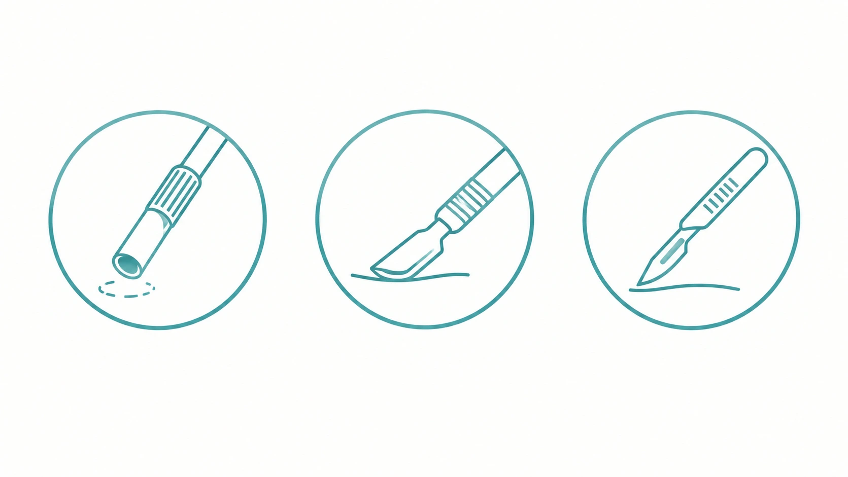
- Suspicious pigmented growths: These are typically sampled by removing the entire visible growth with a small surrounding margin, which gives the clearest possible view of the full structure.
- Deeper or inflammatory-type growths: A small, full-thickness cylindrical sample is often used, since it captures all the skin layers needed for an accurate read.
- Raised, clearly benign-appearing growths: A thin, superficial sampling technique is often sufficient, since these growths generally don’t extend deep beneath the surface.
- Timing matters too: For certain inflammatory skin conditions, sampling at a very specific point in the growth’s evolution – not too early, not too late – genuinely improves diagnostic accuracy.
Treatment: What Skin Growth Removal Actually Involves
Skin growth removal typically begins with numbing the treatment area using a local anesthetic, often combined with a vasoconstricting additive that keeps the surgical field clear and reduces bleeding. This combination has an excellent, well-established safety record even in areas like the fingers, toes, ears, and nose – older concerns about using it in these areas have been firmly addressed by modern clinical evidence. After the numbing medication is given, a short waiting period is observed before the procedure begins, since the area needs a few extra minutes beyond simple numbness to reach its most bloodless state.
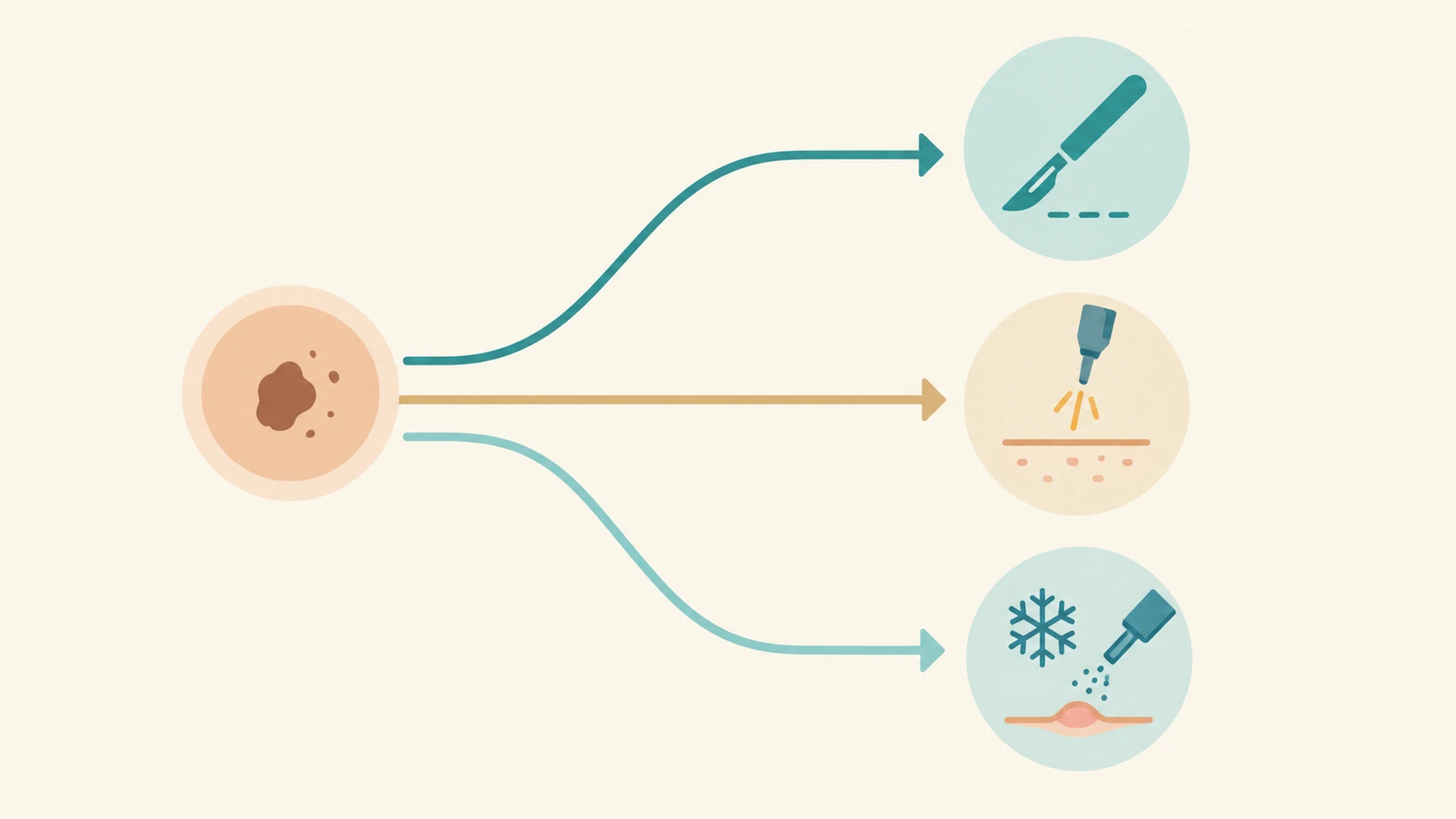
For many common benign growths like skin tags, corns, calluses, and warts, removal is relatively straightforward and completed in a single, quick in-office visit. For other growths, the dermatologist may use a precise scraping-and-cauterizing technique, which physically removes the growth and then uses heat to clear the base and control bleeding – this approach works well for superficial, low-risk, non-facial growths but isn’t used in cases where the full extent of the growth needs to be clearly confirmed. When a growth requires the most thorough possible removal, particularly on the face or in cases needing maximum precision, a meticulous margin-checking technique is used, where the removed tissue is examined in its entirety before the procedure is considered complete.
Removal Method Guide: Matching the Technique to the Growth
| Growth Type | Removal Approach | Why This Approach |
|---|---|---|
| Skin tags, warts, corns, calluses | Simple, direct in-office removal | Clearly benign, well-defined, low recurrence risk |
| Raised, clearly benign growths | Straightforward excision or scraping technique | Shallow depth makes simple removal effective |
| Growths needing full margin confirmation | Meticulous margin-checking technique | Ensures the entire growth has genuinely been cleared |
| Superficial, low-risk, non-facial growths | Scraping-and-cauterizing technique | Efficient, though no tissue sample is preserved for further checking |
| Growths in high-precision or facial zones | Highly thorough, layer-by-layer margin technique | Maximizes healthy tissue preservation in delicate areas |
The right technique depends on what the growth actually turns out to be and where it’s located – this is a decision made with a dermatologist, not a one-size-fits-all approach.
What the Research Shows?
Published dermatologic research supports several specific points about skin growth evaluation and removal:
- Structured magnified examination improves accuracy: Research on specialized magnified skin examination confirms that evaluating specific visual patterns meaningfully improves the ability to identify growths that need closer attention, beyond what visual inspection alone can achieve.
- Numbing medication with vasoconstrictors is safe in extremities: Large clinical series specifically studying this approach in fingers and other extremities report no cases of tissue damage, overturning decades of outdated caution.
- Sampling technique directly affects diagnostic accuracy: Research confirms that choosing the correct tissue sampling method and timing for a given growth type significantly improves the reliability of the resulting diagnosis.
- Thorough margin-checking techniques improve long-term outcomes for certain growths: Published comparisons show that more thorough, complete margin-examination techniques achieve meaningfully better long-term clearance rates than standard sampling methods for growths where complete removal is critical.
These findings reflect published dermatologic research; the right evaluation and removal approach for an individual still depends on an in-person assessment.
Doctor Review Block
This article has been medically reviewed by Dr. Tejansu Dalal, MD (Dermatology), for clinical accuracy. Safe skin growth removal starts well before the procedure itself – a careful, structured evaluation is what correctly separates a growth that needs a simple in-office removal from one that needs a closer, more deliberate approach.
References & Citations
This article draws on dermatologic surgical research, including findings published by the National Center for Biotechnology Information on surgical approaches to skin growth management, and reference material from the National Institutes of Health on the safety of local anesthesia techniques used during removal procedures. Clinical evaluation and safety details reflect published dermatologic research.